
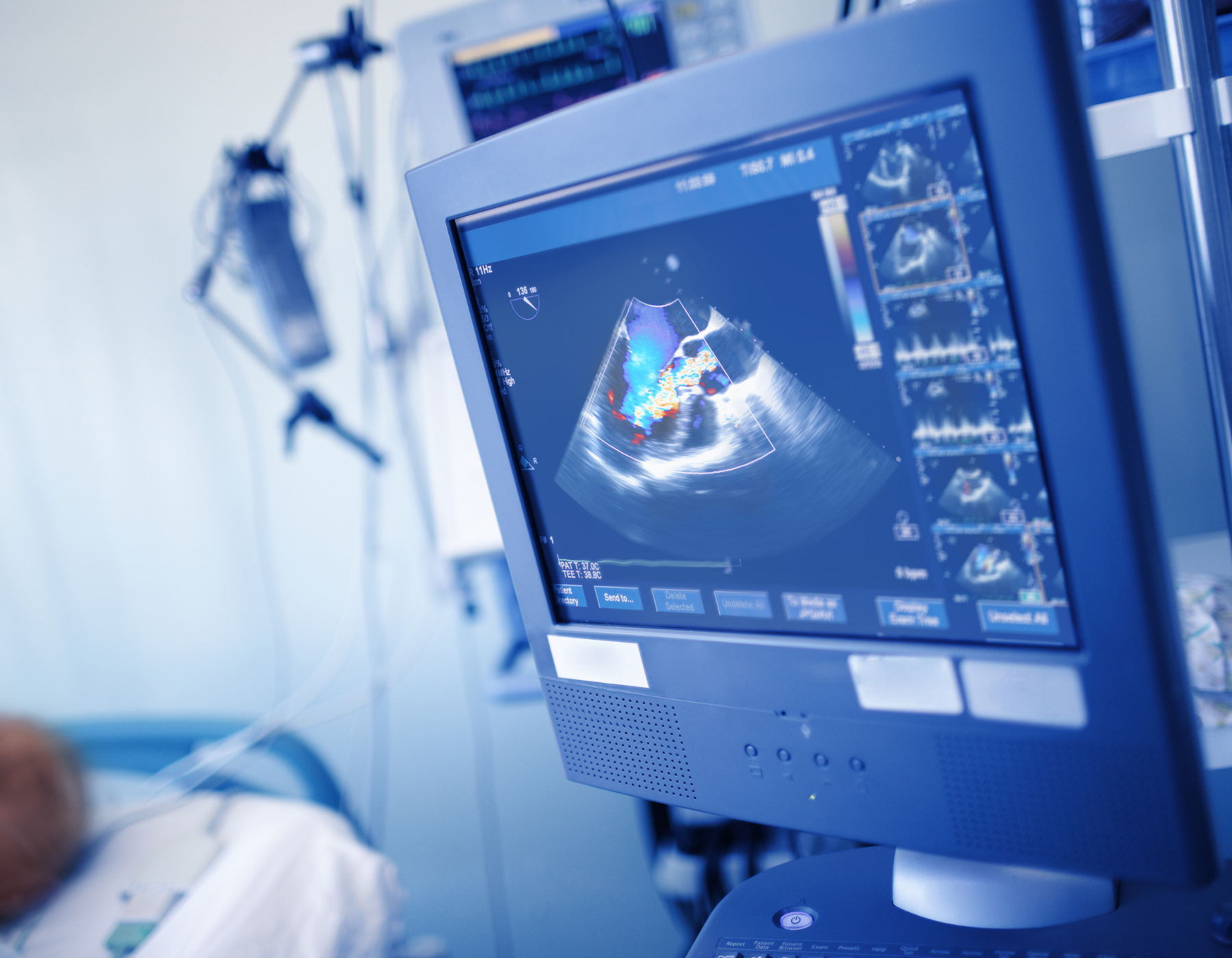
As a medical imaging professional, you know that DICOM compliance is an important topic when it comes to selecting your imaging equipment. We have created a simple guide that will help you understand this set of quality control standards that apply to all medical monitors in the United States.
DICOM stands for Digital Imaging and Communications in Medicine, and it’s an international, nonproprietary protocol created to set a standard in the management and transmission of medical images that include ultrasound, X-Rays, MRIs, CTs, etc., and the patients’ data used in healthcare facilities.
Said standards are divided into five different functions in medical imaging to facilitate its execution, accuracy, and exchange of information between healthcare parties:
- Images and data transmission
- Files retrieving and querying
- Printing or archiving
- Support of digital imaging workflows
- High-quality images for diagnostics
The extension for DICOM files is .dcm and includes the patient data and the image/pixel data. The patient information comes from the EMR/EHR/HIS systems as HL7 data, which gets tightly coupled with the equipment, procedures, and image/pixel data created by the radiology medical imaging devices as DICOM data.
This protocol is a binary Upper-Level Protocol (ULP) over TCP/IP. DICOM's well-known ports are 104, 2761, 2762, and 11112. They are used to process the five functions we mentioned above, from the radiology archival/storage systems like PACS (Picture Archiving and Communication System) and RIS (Radiological Information System) to the workstation for the radiologist.
An example of how the DICOM network protocol architecture looks like:
Network ⇒ TCP/IP ⇒ DICOM ULP for TCP/IP ⇒ UL Service boundary ⇒ DICOM Message Exchange ⇒ Medical Imaging Application.
DICOM not only facilitates the exchange of information in terms of the method, but it also defines the formats professionals in the field of medical imaging should use to ensure consistent files in the healthcare facilities in the United States and the rest of the world that make them easy to read. This way, the interoperability between hardware and software from different equipment, brands, and manufacturers is possible.
Thanks to this functionality, DICOM has become the primary option in the medical imaging world. Most digital medical imaging systems support and comply with the DICOM standard. Additionally, public and private medical institutions have widely accepted it and adopted it as the default option.
DICOM’s History
The Digital Imaging and Communications in Medicine was initially created by the National Electrical Manufacturers Association (NEMA) and the American College of Radiology (ACR) as an initiative to create a functional and standardized way to boost collaboration between professionals in the area of medical imaging.
This project started in the 1980s because it was hard for radiologists to decode the images generated by the machines, and the NEMA and ACR decided to form a standard committee in 1983. Two years later, in 1985, they launched the first version, but soon they realized that it needed improvements. In 1988, the second version was released and gained acceptance among vendors and was then presented at the annual meeting of the Radiological Society of North America (RSNA) in 1990, where new improvements were discovered.
After this, the first large-scale deployment of ACR/NEMA technology was developed in 1992 by the US Army and Air Force as part of the MDIS (Medical Diagnostic Imaging Support), and the following year in 1993, the third version was launched. The name assigned for the project was Digital Imaging and Communications in Medicine, abbreviated DICOM 3.0 as continuity for past versions.
DICOM Part 14
While other parts of the DICOM Standard specify how digital image data can be moved from system to system, it doesn't specify how the pixel values should be interpreted or displayed. DICOM Part 14 provides specifications about a function that relates pixel values to displayed luminance levels to guarantee a reliable image.
When it comes to medical imaging, consistency is key when the digital image appears regardless of where it's being shown; whether it's on the display monitor or as a film on a lightbox, the image must be accurate. Since there wasn't any standard that could regulate how these images were visually presented on any device, DICOM developed a Grayscale Standard Display Function (DICOM Part 14) to ensure an objective, quantitative mechanism for mapping digital images values into a given range of luminance.

The Grayscale Standard Display Function is based on human contrast sensitivity. However, this characteristic is distinctly non-linear within the luminance range of the Grayscale Standard Display Function. Since the human eye is less sensitive in dark areas, this variation in sensitivity facilitates perceiving small changes in luminance in the bright areas of an image than in the dark areas of the image.
By providing a standard in the grayscale display, professionals can distinguish different shades on the target using luminance to make those variations even more visible. Using a luminance range that goes from 0.05 to 4000 cd/m2, the Grayscale Standard Display Function makes it possible to obtain the same accuracy on cathode-ray-tube (CRT) monitors and bright lightboxes used for interpreting x-ray mammography.
Why DICOM is Relevant
DICOM is the global standard used to store, exchange and transfer medical images. These functions have been beneficial for the healthcare industry and the patients because they make sharing information easier, increasing collaboration regardless of professionals' location.
Without a standardized system, it would be hard to share patient data from different devices because each would need its own format and software to translate the information for other equipment. DICOM improves the workflow making it more efficient for physicians, thereby providing patients with a better experience and care.
Traditional image formats like JPEG and TIFF do not allow storing patient information or procedure information, thus they are not an option for diagnostic purposes. On the other hand, DICOM includes relevant information about the patient, the examination, the equipment used, the medical practitioners and equipment operators involved, and the scope of the examination.
When it comes to medical equipment, ERI offers the ultimate technology in hardware and software to give facilities like yours the tools to provide your patients with excellent care and your staff with an optimized workflow that makes their job easier without compromising the quality and accuracy of exams and procedures.
Discover our wide range of DICOM-compliant medical displays that include mammography monitors, radiology displays, surgical monitors, and clinical review displays.