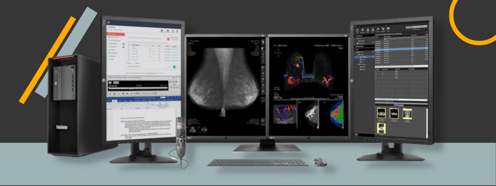
Consistency is key when it comes to medical imaging. From x-rays to CT scans, MRIs to ultrasounds, ensuring that medical images are accurate is paramount to making critical diagnoses and providing effective treatments. In this blog, we'll explore how to achieve consistent presentation of medical images and why this consistency is so crucial for healthcare professionals.
In modern radiology departments, the proper functioning of the image display system is a critical component of a successful information technology platform. Despite the prevalence of advanced computing and high-quality medical display monitors, medical images may be suboptimal for diagnostic purposes if the displays are not configured and maintained correctly.
It is essential that practicing radiologists, physicians, and other healthcare professionals recognize the role of display technology and usage in ensuring high-quality radiology practices.
With advancements in medical technology, healthcare facilities process an increasing amount of imaging data. To improve diagnostic accuracy, medical professionals are using both monochrome and color images. These types of images should be displayed separately, using different grayscale standards.
According to EIZO, a medical monitor manufacturer, in their article How to Accurately Display Both Monochrome and Color Images with the Ideal Grayscale, DICOM Part 14 Grayscale Standard Display Function (GSDF) is suggested for monochrome images, while Gamma 2.2 is recommended for color images. Failure to use the appropriate grayscale could result in lower image quality and potentially lead to missing crucial diagnostic information.
Types of Medical Images Depending on the Source
In the article Consistent Presentation of Medical Images, the author explained that medical practices use two main categories of patient images:
1. Radiology Images: They utilize non-visible energy (such as X-ray, nuclear, ultrasound, or magnetic) to create an interior view of body parts. These types of images exhibit a linear relationship between the image brightness levels and the internal densities of the tissue and bone being scanned.
However, the resulting digital image data shows a logarithmic relationship with the scanned internal densities. To achieve perceptual linearity, darker areas of the scanned images must undergo contrast enhancement when presented on radiology display monitors.
If the logarithmic image data was displayed on a medical display monitor with logarithmic luminance response, the display would produce image brightness levels that correspond more accurately with the density of the scanned object.

2. Medical Video Images: On the other hand, medical video images rely on visible light to form a surface view of body parts or samples, and can be used in applications including endoscopy, surgical, ophthalmology, dermatology, and microscopy.
This modality uses camera-acquired images to visualize surgeries, procedures, and still images captured during examinations in various medical fields, including dermatology and pathology.
These camera-acquired medical images adhere to the 1999 standard from the International Electrotechnical Commission, known as sRGB. This standard was designed to be suitable for brighter viewing environments and is also followed by consumer digital cameras and internet images.
The sRGB luminance response model starts with a linear section just above black with a gamma of 1, then shifts to a power function at higher levels with a gamma of 2.4, averaging a gamma of 2.2 across the entire luminance range.
Because of the differing characteristics involved in scanner-sourced radiology images versus camera-sourced medical video images, different performance requirements are needed for medical grade display monitors used to render the two types of images.
Standards for Radiology Images
DICOM
An image that appears diagnostically useful on one device may lose its value on another. Ensuring that digital images appear the same regardless of the medical display monitor is the primary aim of PS3.14, which is the Grayscale Standard Display Function by DICOM.
DICOM offers a quantitative way of mapping digital image values into a specified range of luminance levels. By providing an application with this relationship between digital values and display luminance, better visual consistency across different devices can be achieved.
This relationship is established based on scientific measurements and models of human perception instead of being dependent on the characteristics of a device or imaging modality or user preferences.
DICOM has gained widespread acceptance within the medical imaging sector, becoming the primary option due to its functionality. It is supported and adhered to by most digital medical imaging systems. Furthermore, both public and private medical institutions have embraced and made it their default option.

ACR – AAPM – SIIM
The ACR-AAPM-SIIM standard refers to a collaborative effort between the American College of Radiology (ACR), the American Association of Physicists in Medicine (AAPM), and the Society for Imaging Informatics in Medicine (SIIM). The primary goal of this standard is to define consistent procedures and protocols for acquiring, transmitting, processing, and archiving medical images.
To achieve this, the standards recommend direct image capture and the use of standardized display protocols, which means that the image data set produced by the modality for diagnostic interpretation is transferred to the image management system with complete spatial resolution (image matrix size) and contrast resolution (pixel bit depth), using the DICOM Standard for image transactions.
This ensures that images can be easily compared and analyzed, regardless of the equipment used or the location of the images. Additionally, the standards emphasize the importance of monitoring and managing equipment performance to maintain consistency and accuracy.
Image display standards are also a critical component of the ACR-AAPM-SIIM standards. The standards recommend using grayscale displays with a minimum luminance of 171 cd/m² and a contrast ratio of at least 250:1. This ensures that images are displayed accurately and can be interpreted correctly by imaging professionals. The standards also specify requirements for color displays and ambient lighting in imaging environments.
Color Images in Radiology
Gamma 2.2 is a specific gamma setting that is commonly used in color grading for brighter viewing conditions, such as in an office setting with bright overhead lighting. As we stated before, Gamma 2.2 is recommended for color images.
Some monitor technologies, such as EIZO’s CAL Switch function, offer preset viewing modes for individual modalities. However, when it comes to viewing both monochrome and color images together, many users prefer sticking to the GSDF mode instead of switching between modes. Here, EIZO addresses this issue with its Hybrid Gamma PXL function.
A study from Kumamoto Chuo Hospital, Japan revealed that 77% of participants found the blended pixel-by-pixel method to be equivalent to Gamma 2.2 when viewing color images. Additionally, 94% of participants reported that grayscale images were displayed with the same quality as GSDF, which is comparable to what users would see on a monochrome monitor.
Luminance in Medical Imaging
In medical imaging, luminance is used to measure the brightness of displays used to view medical images, such as X-rays, CT scans, and MRI scans. To ensure high-quality medical imaging, it is important to have displays with high luminance levels.
For example, according to an article published in the Journal of Medical Imaging in 2018, the American College of Radiology Technical Standards recommends a minimum luminance of 420 cd/m2 for diagnostic X-ray mammography displays.
Luminance plays a crucial role in medical imaging by ensuring the accuracy and quality of medical images. Radiology display monitors used for medical imaging must be equipped with high luminance levels and calibration technology to maintain the grayscale characteristics of images over time.
Calibration of Devices for an Accurate Presentation of Medical Images
Calibration of devices for an accurate presentation of medical images is crucial for ensuring the diagnostic value of medical images remains consistent, regardless of the display device used.
Calibration detects and fixes uncertainties in measurements and brings them within a certain level of accuracy. Calibration of x-ray imaging devices is particularly important because it is key to accurate calibration of the x-ray sources.
Medical display calibration is the first step in the radiology imaging calibration workflow, and it involves measuring the screen luminance. In an article published in 2008 in the Journal of Medical Imaging, the authors explained that photometers are used for calibrating medical imaging grade displays, and they should be accurate to within 5%, except for very low luminance levels.
Additionally, the usage of DICOM calibration software in the medical industry enables consistency in the diagnostic value of medical images. Calibration of instrumentation used for display quality assurance helps to ensure that there are no errors in interpretation. Calibration of devices is done using calibration equipment that has been calibrated to a known accuracy.

Overall, ensuring a consistent presentation of medical images is crucial for accurate diagnosis and effective treatment. The different types of medical images, including those originating from X-rays, CT scans, and MRI scans, all require calibration to ensure that the diagnostic value remains consistent, regardless of the medical display monitor used.
By following these standards and implementing these best practices, healthcare professionals can achieve a consistent presentation of medical images and ensure the highest degree of accuracy and reliability in medical diagnosis and treatment.


